The Tissue Structure Of The Lungs In An Enlarged Model Of Alveoli

Product information:
Product name: Alveolar enlargement model
Product size: 26*15*35cm
Applicable scene: teaching
The Tissue Structure Of The Lungs In An Enlarged Model Of Alveoli – Detailed Respiratory System Learning Tool
Perfect for Medical Education, Biology Classes, Anatomy Demonstrations, Healthcare Training, and Science Learning
The Tissue Structure Of The Lungs In An Enlarged Model Of Alveoli is designed to provide a clear and detailed representation of the microscopic structures found within the human lungs. This educational model enlarges the alveoli and surrounding lung tissue structures so students and educators can better understand how gas exchange occurs in the respiratory system.
Because alveoli are extremely small structures inside the lungs, they are difficult to study using standard diagrams alone. An enlarged model allows learners to observe the structure of alveoli, surrounding capillaries, and the thin tissue walls that enable oxygen and carbon dioxide exchange.
This anatomical teaching model helps simplify complex respiratory concepts by providing a three-dimensional visual representation of lung tissue. It is commonly used in classrooms, medical training environments, laboratories, and healthcare education programs.
Description – The Tissue Structure Of The Lungs In An Enlarged Model Of Alveoli
The Tissue Structure Of The Lungs In An Enlarged Model Of Alveoli is an educational anatomy model designed to illustrate the microscopic structures responsible for gas exchange in the lungs. The model magnifies the alveoli and surrounding tissue so learners can study the detailed structure that supports breathing and oxygen circulation in the human body.
Alveoli are tiny air sacs located at the ends of the bronchioles within the lungs. Their primary function is to allow oxygen from inhaled air to enter the bloodstream while removing carbon dioxide from the body.
Because alveoli are extremely small and numerous, their structure is often difficult to visualize in standard anatomical diagrams. An enlarged educational model provides a clearer representation that helps students understand how these structures function.
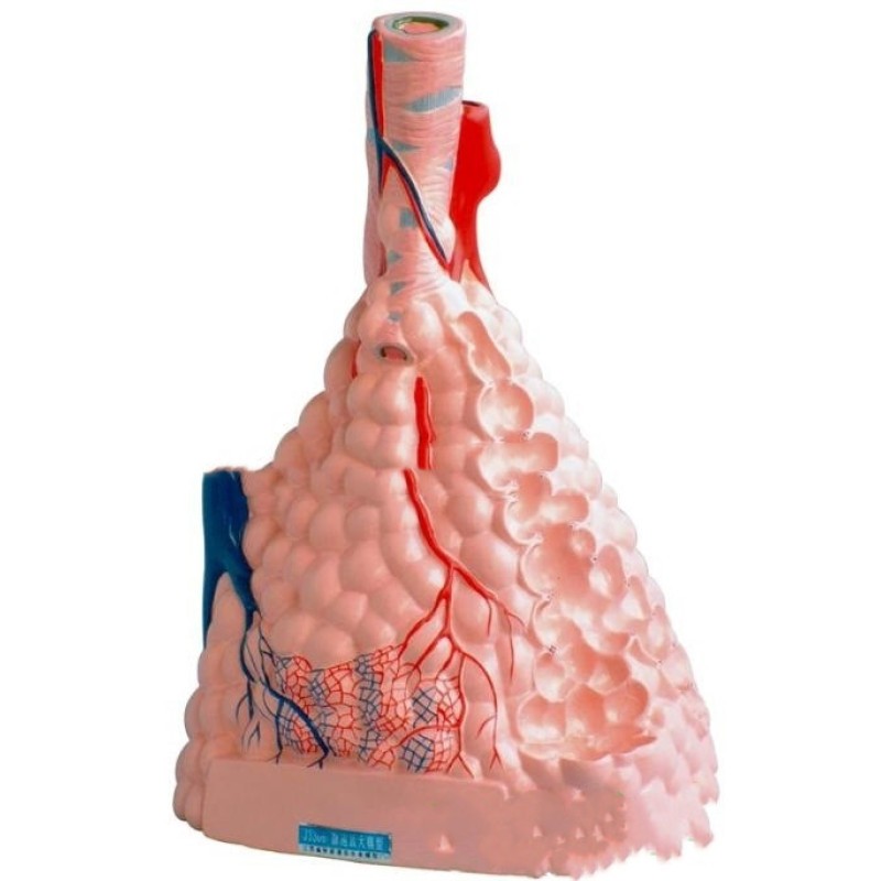
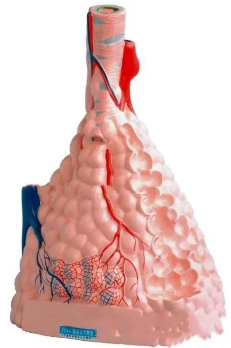
The model typically displays the alveolar sacs, which appear as clusters of small bubble-like structures connected to the bronchioles. These sacs represent the final stage of the respiratory airway where gas exchange occurs.
Surrounding the alveoli are capillary networks, which carry blood close to the thin alveolar walls. This proximity allows oxygen molecules to pass from the air inside the alveoli into the bloodstream.
The model also highlights the thin epithelial tissue layers that form the walls of the alveoli. These delicate tissues are designed to allow efficient gas diffusion between air and blood.
Many enlarged alveoli models use color coding to distinguish different structures within the lung tissue. This visual separation helps students easily identify alveolar sacs, blood vessels, bronchioles, and connective tissue.
Because the model is three-dimensional, learners can examine the alveoli from multiple angles. This improves comprehension compared to flat diagrams found in textbooks.
The model is commonly mounted on a stable base, allowing it to be displayed during lectures, laboratory sessions, or classroom demonstrations.
Medical students often use such models to better understand respiratory physiology, particularly the process of oxygen exchange and carbon dioxide removal.
Another advantage of this educational model is its ability to support interactive teaching methods. Instructors can use the model to demonstrate how air travels through the respiratory tract before reaching the alveoli.
Healthcare educators may also use the model when explaining respiratory conditions or lung function to patients or trainees.
Durable materials are typically used in the construction of anatomical models to ensure they remain suitable for repeated educational use.
Because the model enlarges microscopic structures, it allows learners to clearly observe anatomical features that are otherwise too small to see with the naked eye.
Understanding alveoli is essential for studying respiratory biology, as these tiny sacs play a crucial role in supplying oxygen to the body’s tissues.
The Tissue Structure Of The Lungs In An Enlarged Model Of Alveoli helps bridge the gap between theoretical study and visual understanding by providing a tangible representation of lung tissue structure.
Key Points
Enlarged Alveoli Structure
Magnifies tiny lung air sacs to improve anatomical understanding.
Three-Dimensional Educational Model
Allows learners to view respiratory structures from multiple angles.
Color-Coded Anatomy Design
Helps identify alveoli, capillaries, and surrounding tissues.
Stable Classroom Display Model
Mounted on a base for easy demonstration during lectures.
Benefits
Improves Respiratory System Understanding
Helps students visualize the microscopic structures of the lungs.
Supports Medical and Biology Education
Useful for teaching respiratory anatomy and physiology.
Enhances Interactive Learning
Allows instructors to demonstrate lung function clearly.
Reinforces Visual Memory
3D models help improve retention compared to flat diagrams.
Why Choose This Tissue Structure Of The Lungs In An Enlarged Model Of Alveoli?
The Tissue Structure Of The Lungs In An Enlarged Model Of Alveoli provides a clear and accurate educational representation of one of the most important microscopic structures in the respiratory system.
Its enlarged design allows learners to observe the structure of alveoli, capillaries, and lung tissue in detail. This helps students better understand how oxygen exchange occurs within the lungs.
For educators, medical professionals, and students studying human anatomy, this model provides a valuable visual learning tool that simplifies complex respiratory concepts.
Conclusion
The Tissue Structure Of The Lungs In An Enlarged Model Of Alveoli is an educational anatomy model designed to illustrate the detailed structure of lung tissue and alveoli. By magnifying microscopic respiratory structures, it helps learners better understand the process of gas exchange and lung function.
With its three-dimensional design, clear anatomical representation, and classroom display base, the model serves as a useful tool for medical education, biology teaching, and healthcare training.
The product may be provided by a different brand of comparable quality.
The actual product may vary slightly from the image shown.
Shop amazing plants at The Node – a top destination for plant lovers

.png)


.jpg)


.jpeg)


.jpeg)


.jpeg)


.jpeg)


.jpeg)

.jpeg)

.jpeg)
.jpeg)


.jpeg)
.jpg)

.jpeg)


.jpeg)
.jpeg)


.jpeg)


.jpeg)

.jpeg)

.jpeg)

.jpeg)
.jpeg)


.jpeg)
.jpeg)
.jpeg)


.jpeg)


.jpeg)


.jpg)
.jpeg)


.jpg)


.jpeg)


.png)


.png)