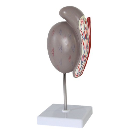
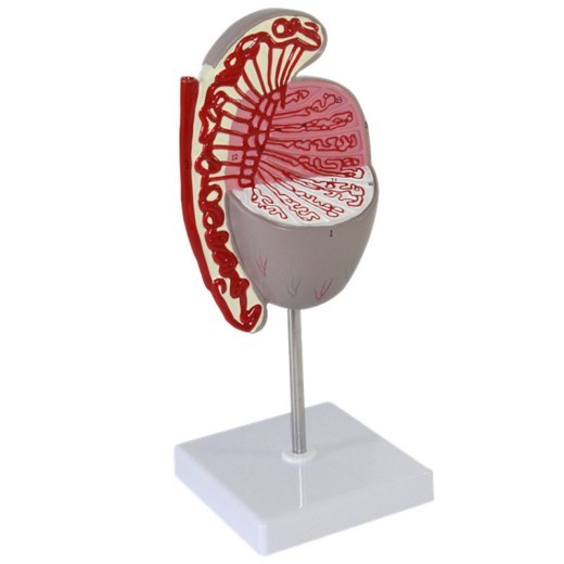
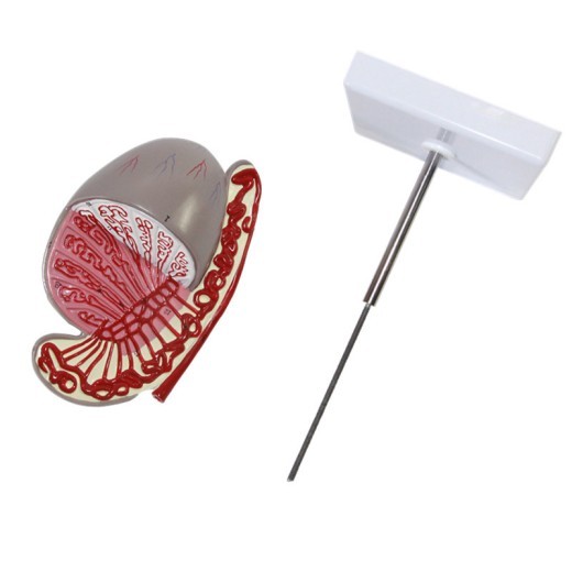
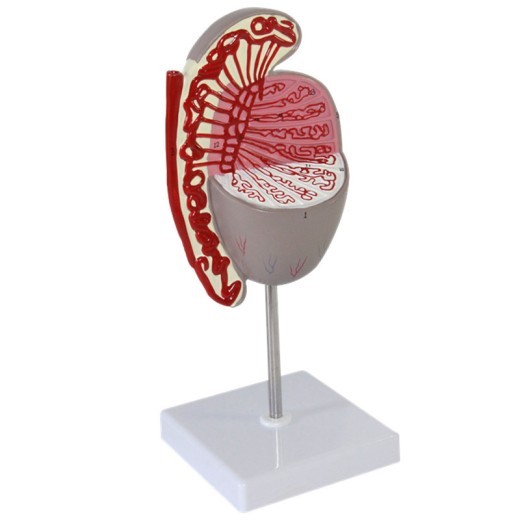
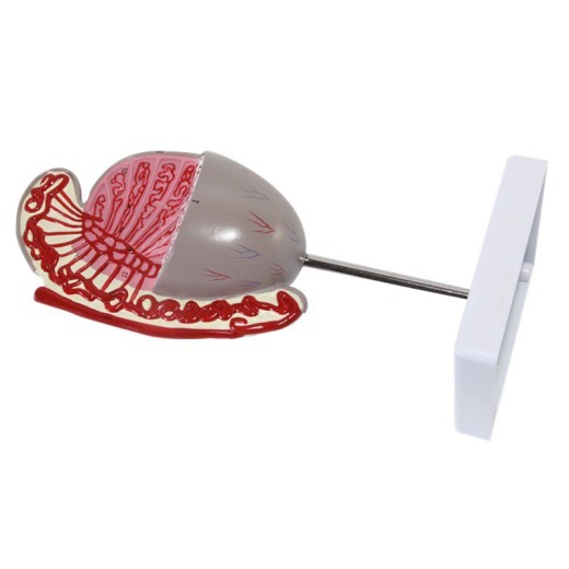
Testicular Model Testicular Structure Guidance Model
Approx $87.38 USD
Introduction to the Testicular Structure Guidance Model
The Testicular Structure Guidance Model is an educational tool designed to aid in the understanding of the male reproductive system, specifically focusing on testicular anatomy. It provides a detailed view of the structures within the testes, making it invaluable for students, educators, healthcare professionals, and reproductive health trainers. With accurate detailing and high-quality construction, this model is perfect for classrooms, clinics, and laboratories in New Zealand, offering an effective way to study testicular function, sperm production, and other related concepts. By giving a tangible representation of complex anatomical details, this model enhances learning and comprehension for those studying human biology and reproductive health.
H2: Key Features of the Testicular Structure Guidance Model
1. Realistic Detailing of Testicular Anatomy
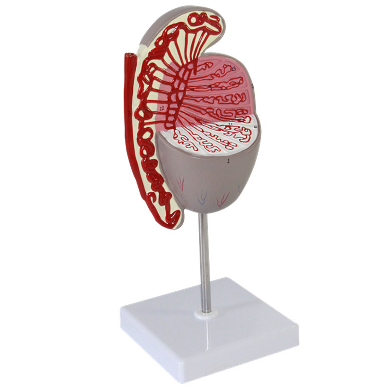
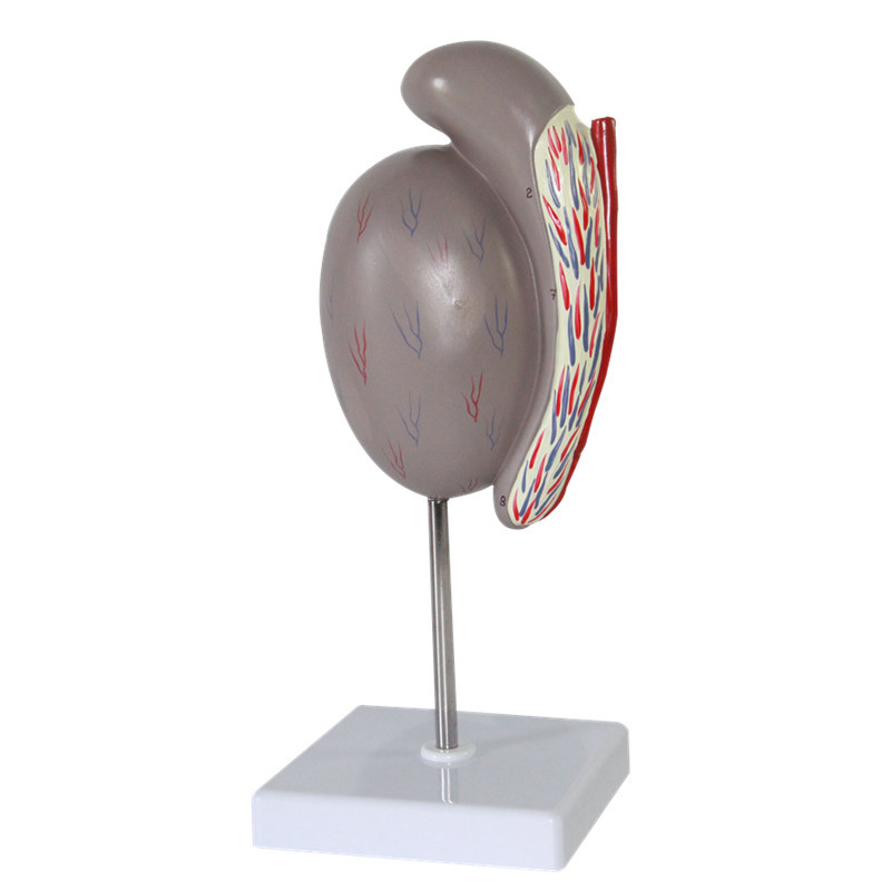
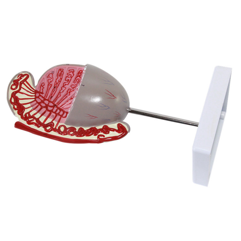
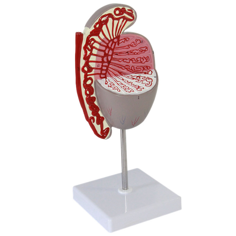
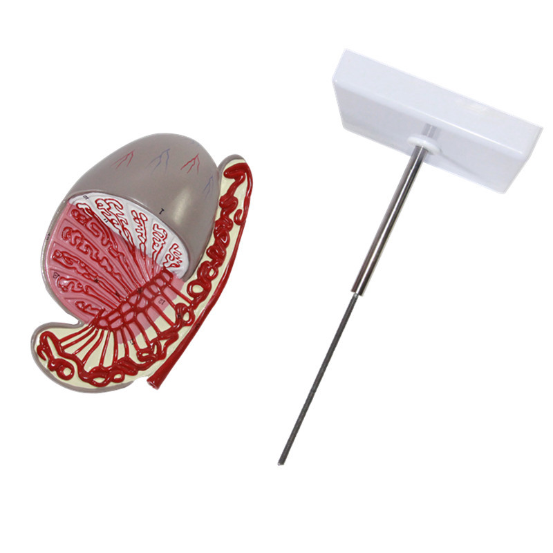
The Testicular Structure Guidance Model accurately represents the structures within the testes, including the seminiferous tubules, epididymis, vas deferens, and surrounding blood vessels. Each part is designed to provide a clear view of how sperm production occurs, helping students and trainees understand the role of each component in male reproduction. The intricate detailing allows users to appreciate the complexity of testicular anatomy, making it ideal for educational use.
2. Durable and High-Quality Materials
Constructed from high-quality, non-toxic materials, this model is built to withstand frequent handling and use in educational settings. Its durability ensures that it remains intact even with regular use, making it suitable for classrooms, laboratories, and healthcare environments. This long-lasting construction allows students and educators to engage with the model over time, providing a reliable resource for ongoing learning.
3. Color-Coded and Labeled Structures
To aid in learning, the model features color-coded and labeled parts, highlighting the main areas of testicular anatomy. This includes the seminiferous tubules, epididymis, and blood vessels, among others. The color-coded sections make it easier to distinguish between different structures, while labels help guide users to quickly identify each part. This system is particularly beneficial for visual learners, enhancing their ability to retain information.
4. Cross-Sectional View for In-Depth Study
This model provides a cross-sectional view of the testes, allowing users to examine the internal structures in detail. This design enables a better understanding of the layers within the testicle and offers a visual guide for studying complex processes such as spermatogenesis. The cross-sectional view allows for an in-depth examination of each structure, making it a valuable tool for advanced learning in biology and health sciences.
5. Portable and Display-Friendly Size
Compact and easy to transport, the Testicular Structure Guidance Model is designed to fit well in various educational and professional settings. Its portable size allows it to be easily moved between classrooms, labs, and clinics, offering flexibility for different teaching scenarios. The model also features a stable base, making it suitable for display on desks, shelves, or in healthcare settings where it can serve as a useful reference for patient education.
6. Gift-Ready Packaging for Academic and Professional Use
The model comes packaged in a secure and presentation-ready box, making it an ideal gift for students, healthcare professionals, or educators focusing on male reproductive health. The packaging provides protection during transit, ensuring the model arrives in perfect condition. This gift-ready option is particularly suitable for graduation gifts, academic awards, or as a resource for beginning medical training.
H2: Why Choose the Testicular Structure Guidance Model?
1. Ideal for Reproductive Health Education
The Testicular Structure Guidance Model is an essential tool for teaching male reproductive anatomy, offering students and trainees a hands-on way to understand testicular structures. By providing a realistic representation of testicular anatomy, this model aids in lectures, discussions, and practical lessons on the male reproductive system, helping students grasp complex concepts related to male fertility and health.
2. Suitable for Medical Training and Patient Education
Beyond academic use, this model is also valuable in medical training programs and patient education. Medical professionals can use the model to explain testicular health, sperm production, and the impact of various conditions on male reproductive health. For patients, the model offers a visual aid to understand their own anatomy better, enhancing communication between healthcare providers and patients regarding topics such as testicular self-exams, fertility, and medical procedures.
3. Supports Visual and Kinesthetic Learning
This model caters to both visual and kinesthetic learners, making it an engaging tool for a wide range of students. The color-coded sections provide visual cues that enhance understanding, while the tactile nature of the model allows students to physically explore each part, helping to reinforce theoretical knowledge. For students who benefit from hands-on learning, the Testicular Structure Guidance Model offers a practical and memorable approach to studying anatomy.
4. Useful for Exam Preparation and Advanced Studies
Students preparing for exams or advancing in their studies can benefit greatly from this model. Its realistic detailing and labeled sections make it an excellent study aid, helping students review and memorize key parts of testicular anatomy. For advanced learners, the model provides an in-depth view of the seminiferous tubules, spermatogenesis, and hormonal interactions, reinforcing their understanding of male reproductive biology.
5. Educational Display for Clinics and Classrooms
The Testicular Structure Guidance Model is a valuable display piece for both educational and clinical environments. In classrooms, it enhances lessons on reproductive health, while in clinical settings, it can serve as a reference for consultations and patient discussions. For New Zealand educators and healthcare providers, this model is a meaningful way to promote awareness and understanding of male reproductive health.
H2: Maintenance and Care Tips for Your Testicular Structure Guidance Model
To keep your Testicular Structure Guidance Model in excellent condition, follow these care tips:
-
Dust Regularly: Use a soft, dry cloth or small brush to dust the model, particularly around the labeled and detailed areas.
This will help keep it clean and maintain its vibrant colors.
-
Avoid Direct Sunlight: Prolonged exposure to direct sunlight can fade colors. Place the model in a shaded area or a display
case to prevent any fading over time.
-
Handle with Care: While durable, the model’s detailed sections require gentle handling to prevent breakage or damage to
labels. Avoid applying excessive pressure to small or delicate parts.
- Store Safely: If you need to transport or store the model, use its original packaging or a protective container to prevent any potential damage. Proper storage helps maintain its quality for future use.
Material information:
Material: pvc
Specification: JY/15103
Category: Visceral Anatomy Series
Packing list:
Testis model*1
Size Information:
27*11*11cm
Product information:
The product may be provided by a different brand of comparable quality.
The actual product may vary slightly from the image shown.
Shop amazing plants at The Node – a top destination for plant lovers



.jpg)




.jpg)




.jpeg)


.jpeg)


.jpeg)


.jpeg)


.jpeg)

.jpeg)

.jpeg)

.jpeg)


.jpeg)
.jpg)

.jpeg)


.jpeg)
.jpeg)


.jpeg)


.jpeg)


.jpeg)

.jpeg)

.jpeg)

.jpeg)





.jpeg)
.jpeg)
.jpeg)





.jpeg)



.jpeg)





.jpg)
.jpeg)



.jpg)


ulva-Logo.jpg)


.jpeg)


.png)









.png)
