One-To-One Medical Simulation Human Skull Model

Product information:
Product material: imported environmentally friendly PVC material
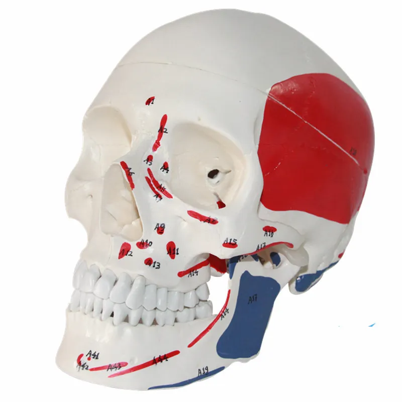
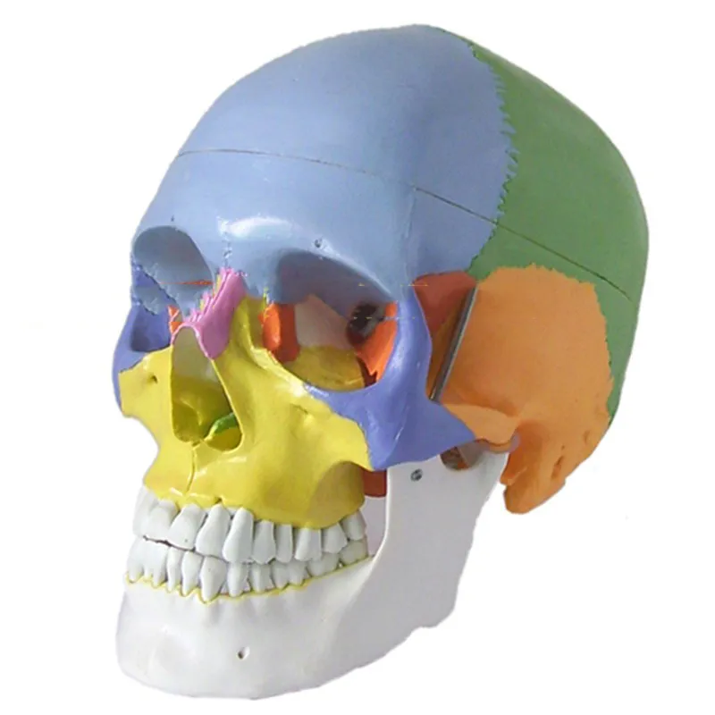
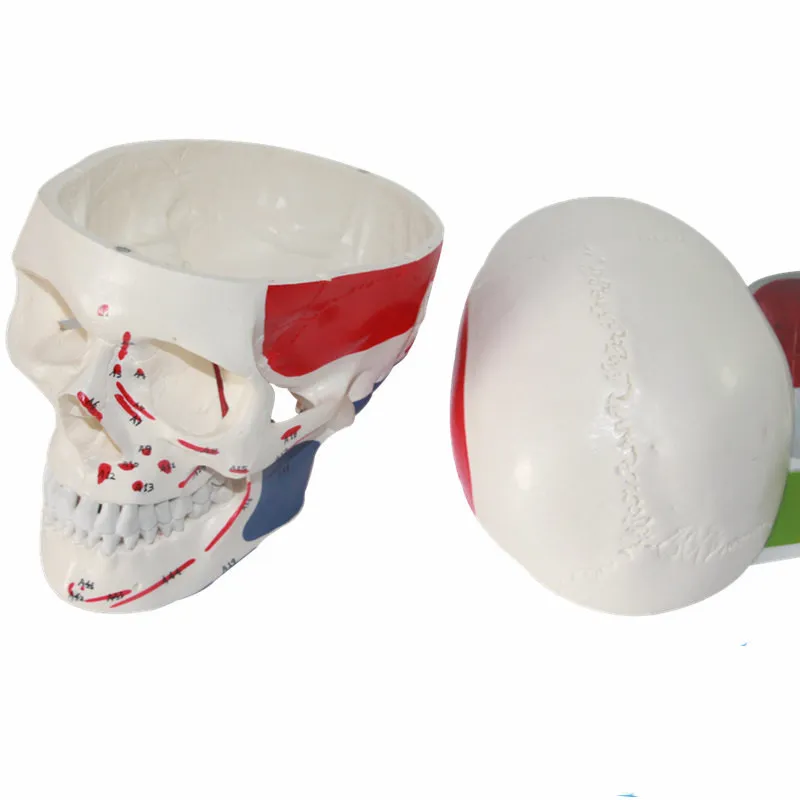
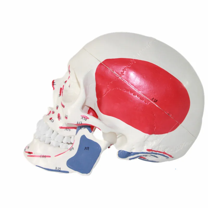
Basic features: Highly simulatedStyle A: Muscle Coloring Skull
B style: pure white skull (no digital logo)
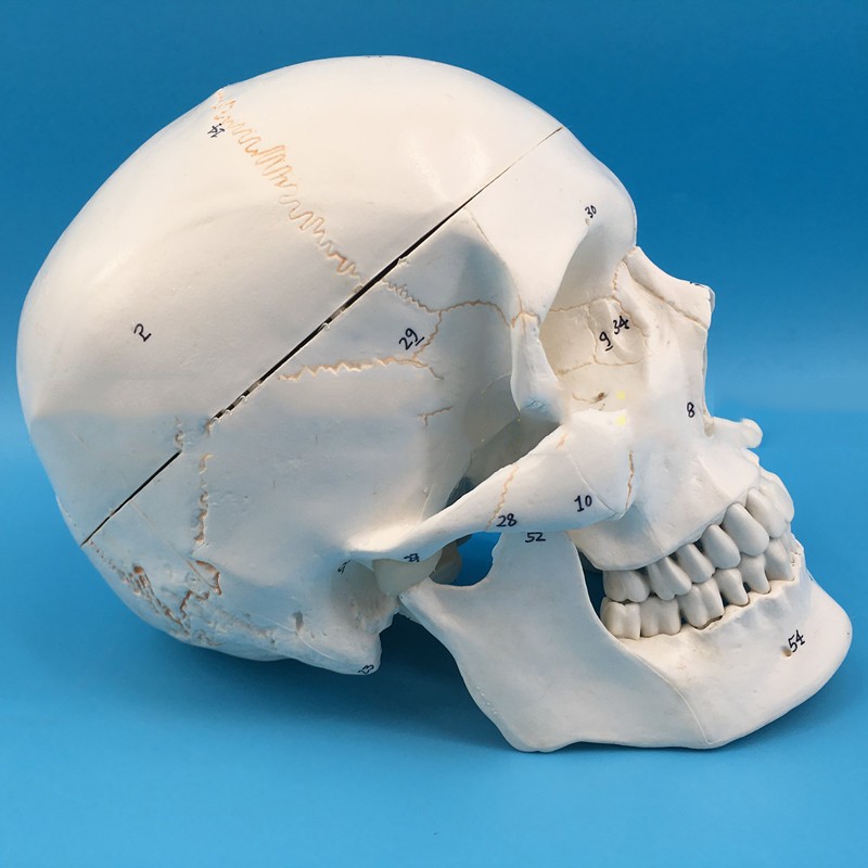
Style C: Skull with sutures (with digital code)
D style: colorful skull (with instruction card)
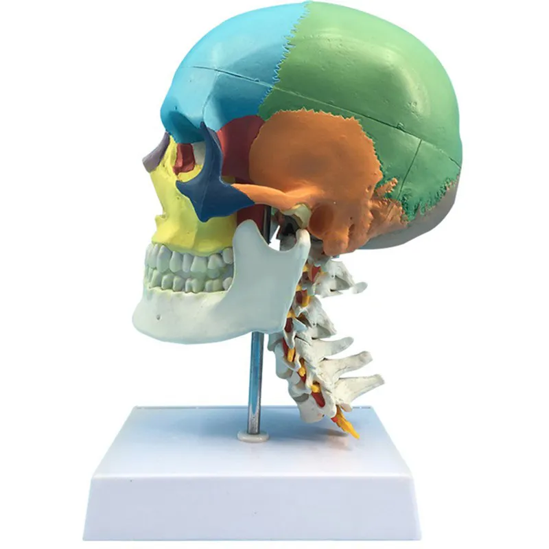
E style: colorful skull with cervical spine


One-To-One Medical Simulation Human Skull Model – Life-Size Anatomical Study Tool
Perfect for Medical Education, Anatomy Classes, Healthcare Training, and Biology Study
The One-To-One Medical Simulation Human Skull Model is designed to provide a detailed life-size representation of the human skull for educational and medical training purposes. Anatomical models like this allow students, educators, and healthcare professionals to study the complex structure of the skull in a clear and interactive way. Understanding the skull is essential in many areas of medical education, including anatomy, dentistry, surgery, and physical therapy. Because the skull contains numerous bones, cavities, and structural features, a physical model helps learners visualize and understand these elements more effectively than flat diagrams alone. This one-to-one scale model replicates the natural size and proportions of the human skull, making it suitable for classroom demonstrations, laboratory study, and medical training environments.
Description – One-To-One Medical Simulation Human Skull Model
The One-To-One Medical Simulation Human Skull Model is an anatomical educational model designed to accurately represent the structure of the human skull. Built to a full one-to-one scale, the model reflects the natural proportions of a real human skull, allowing learners to examine its anatomical features closely. The skull plays an important role in protecting the brain and supporting facial structure. It is composed of multiple bones that fit together to form the cranium and facial skeleton. This model displays these bones in a detailed and organized way, helping learners understand how the skull is constructed. One of the key features of the model is its life-size accuracy. Because the model is built to the same scale as a real skull, it allows students to study the spatial relationships between bones and anatomical structures. The model typically highlights important areas such as the cranial cavity, which houses the brain, and the facial bones, which form the structure of the face. Other visible structures may include the eye sockets (orbital cavities), nasal cavity, jaw structure, and various openings that allow nerves and blood vessels to pass through the skull. Many educational skull models are designed with removable or articulated components, such as a movable jaw. This allows learners to observe how the lower jaw moves in relation to the rest of the skull. Being able to open and close the jaw provides insight into the mechanics of chewing and speaking. The model is typically constructed from durable materials that replicate the appearance and texture of bone. This durability ensures that the model can withstand repeated handling in classrooms or laboratories. The detailed surface structure of the skull helps learners identify bone landmarks, sutures, and other anatomical features that are important for medical studies. Medical students often use skull models to study cranial anatomy, which is essential for understanding head injuries, neurological conditions, and surgical procedures. Dentistry students also benefit from studying skull models because they provide insight into the relationship between the jaw, teeth, and surrounding bone structures. Another advantage of the model is its ability to support interactive learning. Instructors can point to specific areas of the skull during lectures while students observe the physical structure directly. The model is often mounted on a stable base, allowing it to stand upright during demonstrations or display. In healthcare education settings, anatomical models can also be used to help explain medical concepts to patients or trainees. The One-To-One Medical Simulation Human Skull Model provides a realistic visual reference that helps simplify complex anatomical structures for easier understanding. Because of its realistic design and life-size scale, the model is widely used in medical schools, biology laboratories, healthcare training programs, and classroom environments.
Key Points
Life-Size One-To-One Scale
Represents the natural proportions of a real human skull. Detailed Anatomical Structure
Displays cranial bones, facial bones, and important anatomical landmarks. Movable Jaw Design
Allows demonstration of jaw movement and skull mechanics. Durable Educational Construction
Designed for repeated classroom and training use.
Benefits
Improves Anatomy Learning
Helps students visualize skull structure more clearly. Supports Medical Training
Useful for studying cranial anatomy and facial structures. Enhances Classroom Demonstrations
Allows teachers to explain anatomy with a physical model. Encourages Interactive Learning
Students can observe and examine anatomical features directly.
Why Choose This One-To-One Medical Simulation Human Skull Model?
The One-To-One Medical Simulation Human Skull Model offers an effective educational tool for studying human cranial anatomy. Its life-size design and detailed structure allow learners to explore the skull’s complex features in a clear and interactive way. Unlike flat diagrams, this three-dimensional model provides a physical representation that improves understanding of how bones connect and function within the skull. For educators, students, and healthcare professionals seeking a reliable anatomical study aid, this skull model provides a practical and informative solution.
Conclusion
The One-To-One Medical Simulation Human Skull Model is a life-size anatomical teaching tool designed to illustrate the structure of the human skull. With its realistic proportions, detailed features, and durable construction, it supports effective learning in medical and educational environments. Whether used in classrooms, laboratories, or healthcare training programs, this model helps learners better understand cranial anatomy and the complex structure of the human head.
The product may be provided by a different brand of comparable quality.
The actual product may vary slightly from the image shown.
Shop amazing plants at The Node – a top destination for plant lovers

.png)


.jpg)


.jpeg)


.jpeg)


.jpeg)


.jpeg)


.jpeg)

.jpeg)
.jpeg)
.jpeg)


.jpeg)
.jpg)
.jpeg)


.jpeg)
.jpeg)


.jpeg)


.jpeg)

.jpeg)

.jpeg)

.jpeg)
.jpeg)


.jpeg)
.jpeg)
.jpeg)


.jpeg)


.jpeg)


.jpg)
.jpeg)


.jpg)


.jpeg)


.png)


.png)