Enlarged Lung Alveoli Tissue Structure Model

Overview:
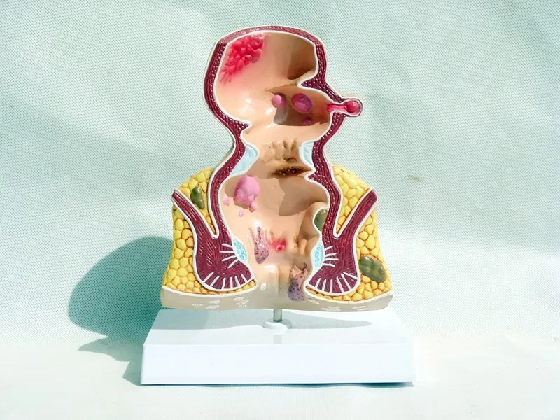
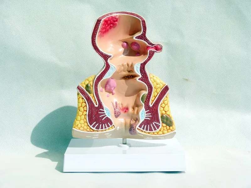
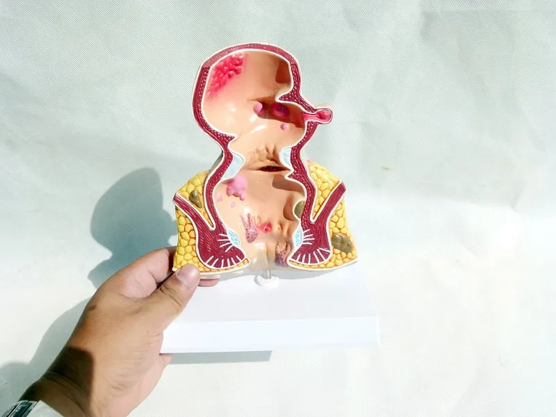
Rectal hemorrhoids lesion model Internal and external anatomy of hemorrhoids Anorectal department Colon and rectum pathology model
Specification:
Material: Plastic
Type: Model
Baby description: Hemorrhoid model rectal lesion model
Features: This model, about 5 times the size, shows various diseases of the rectum and anus. Common anorectal diseases, such as hemorrhoids,
gang fistulas, fissures and 2 types of pus, are shown in very detailed. This model also illustrates the pathological characteristics of
ulcerative colon, xi meat and rectal AI, which is very suitable for teaching and communication between doctors and patients.
Enlarged Lung Alveoli Tissue Structure Model – Detailed Respiratory System Educational Display
Perfect for Medical Education, Biology Classes, Anatomy Demonstrations, Healthcare Training, and Science Learning
The Enlarged Lung Alveoli Tissue Structure Model is designed to provide a clear and highly detailed visual representation of the microscopic structures found within the lungs. By enlarging the alveoli and surrounding tissue structures, this anatomical model helps students and educators understand how the respiratory system performs gas exchange.
Because alveoli are extremely small air sacs located deep within the lungs, they are difficult to study through diagrams alone. This enlarged educational model provides a three-dimensional view of lung tissue, allowing learners to observe how oxygen and carbon dioxide are exchanged between the lungs and bloodstream.
Commonly used in classrooms, medical training environments, laboratories, and health education programs, this anatomical model helps simplify complex respiratory concepts and improve learning experiences.
Description – Enlarged Lung Alveoli Tissue Structure Model
The Enlarged Lung Alveoli Tissue Structure Model is an anatomical teaching aid designed to illustrate the detailed structure of alveoli and surrounding lung tissues. The model magnifies these microscopic respiratory structures so that learners can observe their organization and function more clearly.
Alveoli are tiny air sacs located at the ends of bronchioles within the lungs. Their primary function is to facilitate the exchange of gases—oxygen entering the bloodstream and carbon dioxide being expelled from the body.
Because these structures are extremely small, educational models enlarge them to allow better observation and understanding.
The model typically displays clusters of alveolar sacs, which appear as small bubble-like chambers connected to airway passages. These sacs represent the final stage of the respiratory airway system.
Surrounding the alveoli are capillary networks that carry blood through tiny vessels. These blood vessels run extremely close to the thin alveolar walls, allowing oxygen molecules to pass from the air into the bloodstream.
The model also highlights the thin epithelial tissue layers that form the walls of the alveoli. These tissues are designed to be extremely thin so gases can diffuse easily between air and blood.
Many models include color-coded sections to help distinguish the different structures within lung tissue. These colors allow learners to identify air passages, alveolar sacs, blood vessels, and connective tissue more easily.
The three-dimensional structure of the model allows students to view lung tissue from multiple angles, which improves comprehension compared to flat illustrations.
Mounted on a stable display base, the model can be used during lectures or classroom demonstrations where instructors explain respiratory anatomy and physiology.
Medical students often use these models when studying the respiratory system because they illustrate how air travels through the bronchioles and into the alveoli.
Healthcare educators may also use the model to explain respiratory function or lung conditions to patients or trainees.
The durable construction materials allow the model to be used repeatedly in educational environments without losing structural detail.
Because it magnifies structures that are normally microscopic, the model provides learners with a better understanding of how lung tissues function within the respiratory system.
The Enlarged Lung Alveoli Tissue Structure Model helps bridge the gap between theoretical knowledge and visual understanding by allowing students to observe the anatomical features of lung tissue directly.
Key Points
Enlarged Alveoli Structure
Magnifies tiny lung air sacs for improved anatomical understanding.
Three-Dimensional Educational Model
Allows viewing from multiple angles for detailed study.
Color-Coded Anatomy Design
Different sections highlight lung tissue components clearly.
Stable Display Base
Provides easy classroom demonstration and viewing.
Benefits
Improves Respiratory System Understanding
Helps learners visualize the microscopic structures of the lungs.
Supports Medical and Biology Education
Useful for teaching respiratory anatomy and physiology.
Enhances Classroom Demonstrations
Allows instructors to clearly explain lung function.
Reinforces Visual Memory
Three-dimensional models improve knowledge retention.
Why Choose This Enlarged Lung Alveoli Tissue Structure Model?
The Enlarged Lung Alveoli Tissue Structure Model provides an effective educational tool for studying respiratory anatomy. By enlarging the tiny structures inside the lungs, the model helps learners clearly understand how alveoli function in gas exchange.
Its three-dimensional design allows for interactive demonstrations and improves comprehension of lung physiology.
For educators, students, and healthcare professionals seeking a visual aid for respiratory system education, this anatomical model offers a practical and informative solution.
Conclusion
The Enlarged Lung Alveoli Tissue Structure Model is a detailed educational tool designed to illustrate the microscopic structure of lung tissue and alveoli. By magnifying these structures, the model helps learners better understand how the respiratory system supports oxygen exchange.
With its clear anatomical design and classroom-ready display base, the model serves as a valuable resource for medical training, biology education, and healthcare instruction.
The product may be provided by a different brand of comparable quality.
The actual product may vary slightly from the image shown.
Shop amazing plants at The Node – a top destination for plant lovers

.png)


.jpg)


.jpeg)


.jpeg)


.jpeg)


.jpeg)


.jpeg)

.jpeg)

.jpeg)
.jpeg)


.jpeg)
.jpg)

.jpeg)


.jpeg)
.jpeg)


.jpeg)


.jpeg)

.jpeg)

.jpeg)

.jpeg)
.jpeg)


.jpeg)
.jpeg)
.jpeg)


.jpeg)


.jpeg)


.jpg)
.jpeg)


.jpg)


.jpeg)


.png)


.png)