


Ear Model

Introduction to the Ear Model
The Ear Model is an essential educational tool for those studying human anatomy, particularly the auditory system. With realistic detailing, this model provides an accurate representation of the ear's outer, middle, and inner parts, helping users understand how sound is transmitted and processed. Suitable for students, educators, and healthcare professionals, this model is ideal for classroom lessons, clinics, and training programs across New Zealand. It allows users to explore the ear’s complex structure in three dimensions, making it easier to learn about hearing, balance, and ear health.
H2: Key Features of the Ear Model
1. Detailed Representation of Ear Anatomy
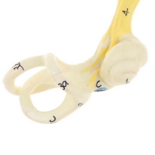
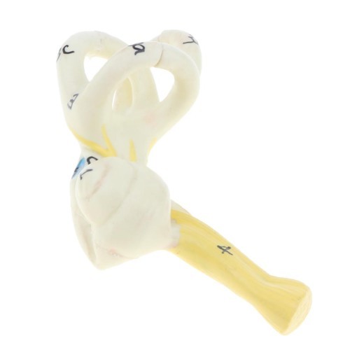
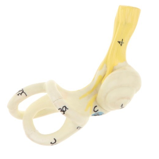
The Ear Model is carefully crafted to display the three main sections of the ear: the outer ear, middle ear, and inner ear. Each part includes accurately scaled components, such as the ear canal, tympanic membrane (eardrum), ossicles (tiny ear bones), cochlea, and semicircular canals. This detailed depiction makes it easy for users to identify each part and understand the pathways through which sound travels.
2. High-Quality and Durable Materials
Constructed from premium, non-toxic materials, this ear model is designed to withstand regular handling in educational and clinical settings. Its durable build ensures that it holds up well over time, making it suitable for anatomy classrooms, ENT (Ear, Nose, Throat) clinics, and audiology training programs. This long-lasting quality provides a valuable resource for ongoing learning and demonstration.
3. Color-Coded Parts for Easy Identification
The Ear Model includes color-coded sections to differentiate between the outer, middle, and inner ear structures. This color-coding helps users quickly identify each area and understand the relationships between the ear’s components. This feature is especially beneficial for visual learners, enhancing comprehension and making it easier to remember the ear’s anatomy.
4. Cross-Sectional View for Enhanced Learning
Some Ear Models offer a cross-sectional design, allowing users to see the internal structures and connections between each part of the ear. This view provides an in-depth look at how sound waves travel through the ear canal, vibrate the eardrum, and move through the ossicles before reaching the cochlea. This cross-sectional view makes complex processes, such as sound transmission and balance, easier to grasp.
5. Compact and Display-Ready
Designed to be compact and display-friendly, the Ear Model fits well on desks, shelves, or medical demonstration areas. It comes with a stable base that keeps it secure during lessons or consultations, making it easy to move between different locations. This portability and stable design make it a versatile tool for classrooms, clinics, and training environments.
6. Perfect for Medical Training and Patient Education
The Ear Model is also an excellent tool for patient education in ENT clinics and audiology centers. Healthcare professionals can use it to explain ear functions, common issues (such as ear infections or hearing loss), and treatment options. The model provides a clear visual aid that makes it easier for patients to understand their ear health, fostering better communication and patient engagement.
H2: Why Choose the Ear Model?
1. Essential for Anatomy and Health Education
The Ear Model is a vital resource for teaching ear anatomy, especially in medical and health science programs. For students learning about the auditory system, it provides a realistic visual representation that connects theoretical knowledge with practical understanding. This model makes anatomy lessons more interactive and accessible, helping students gain a better grasp of how the ear functions.
2. Ideal for ENT and Audiology Training Programs
In ENT and audiology training programs, this model is essential for understanding ear functions, disorders, and treatments. Trainers can use it to illustrate issues such as ear infections, conductive and sensorineural hearing loss, and balance disorders. For New Zealand’s healthcare training programs, this model is an indispensable tool that supports advanced learning in the auditory system and ENT fields.
3. Supports Visual and Kinesthetic Learners
This ear model is particularly valuable for visual and kinesthetic learners, as it allows them to see and touch each structure in the ear. The color-coded parts aid visual identification, while the hands-on nature of the model makes it easier to understand and retain complex auditory anatomy. This interactive approach enhances learning, making it more engaging and effective.
4. Aids in Exam Preparation and Practical Learning
Students preparing for exams or practical assessments will find the Ear Model an invaluable study aid. Its detailed design and labeled parts make it easy to review and memorize the essential structures of the ear. Medical students and trainees can use this model to reinforce their understanding before assessments, ensuring they are well-prepared to identify and explain ear anatomy.
5. Educational Display for Clinics and Classrooms
Beyond its educational function, the Ear Model is an excellent display piece for clinics, classrooms, and training facilities. In clinical settings, it can be used to explain conditions like ear infections, hearing loss, or tinnitus to patients, enhancing their understanding. In classrooms, it creates an engaging environment, providing students with a tangible reference for exploring ear anatomy. For New Zealand educators and healthcare providers, this model is a valuable tool for promoting awareness and understanding of ear health.
H2: Maintenance and Care Tips for Your Ear Model
To keep your Ear Model in excellent condition, follow these care tips:
-
Dust Regularly: Use a soft cloth or brush to dust the model, especially around the detailed areas. Regular cleaning
preserves the model’s appearance and prevents dirt buildup.
-
Avoid Excessive Sunlight: Prolonged exposure to direct sunlight can fade colors over time. Place the model in a shaded area
or indoors to maintain its vibrant appearance.
-
Handle with Care: Although durable, handle the model carefully to avoid damaging delicate parts. Avoid pulling or twisting
small structures, as they may be sensitive to pressure.
- Store Safely When Not in Use: If not displayed, store the model in a safe, dust-free area or in its original packaging to protect it from potential damage. Proper storage helps extend its lifespan for long-term educational use.
-
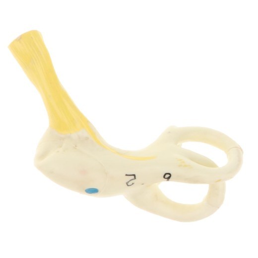
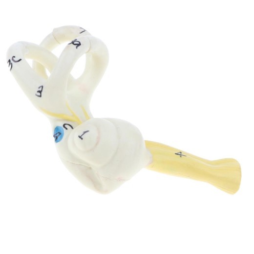
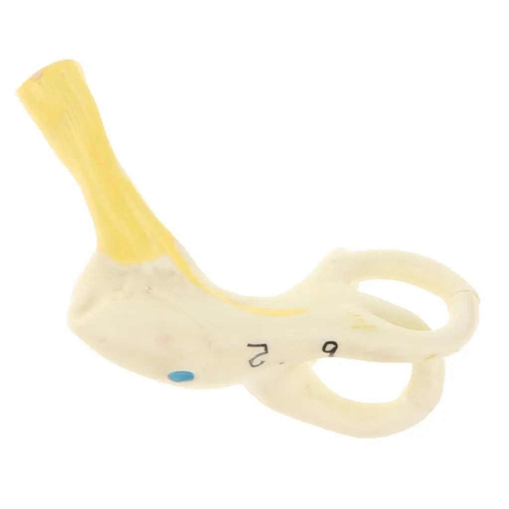
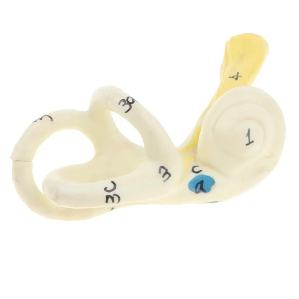
Magnification 5x Human Ear Semicircular Canal Cochlear Model Lab Supplies
Description:
- 100% brand new and with high quality
- Magnification 5x Ear Semicircular Canal Cochlear Model with Labeled Brain Regions for School Medical Study Display
- Help a better understanding of human ear semicircular canal Labyrinth structure
- Great for school teaching tool, learning display, and collectibles, also will be a great addition to your lab supplies
- Material: PVC Material
- Size(LxWxH): Approx. 10 x 7 x 4 cm / 3.94 x 2.76 x 1.57 inch
Package Includes:
1 Enlarged Human Ear Semicircular Canal Cochlear Model
Note:
Due to the manual measurement, please allow 1-5mm differences in size
-


The product may be provided by a different brand of comparable quality.
The actual product may vary slightly from the image shown.
Shop amazing plants at The Node – a top destination for plant lovers
.png)


.jpg)


.jpeg)


.jpeg)


.jpeg)


.jpeg)


.jpeg)

.jpeg)

.jpeg)
.jpeg)


.jpeg)
.jpg)

.jpeg)


.jpeg)
.jpeg)


.jpeg)


.jpeg)
.jpeg)

.jpeg)

.jpeg)
.jpeg)


.jpeg)
.jpeg)
.jpeg)


.jpeg)


.jpeg)


.jpg)
.jpeg)


.jpg)


.jpeg)


.png)


.png)